Eye diseases and conditions
Retinal Tears and Detachments

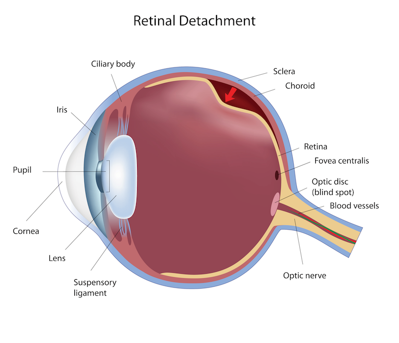
The retina is the layer in the back of your eye that picks up the images you see, much like the film in a camera. A retinal detachment occurs when the retina separates from the back of the eye. This is a serious problem that can cause blindness. Fortunately, there are treatments available, but it important to diagnose it as soon as possible. Retinal detachments will generally come secondary to a retinal tear. Retinal tears most often occur from a vitreous detachment. The vitreous is the clear gel-like material that fills the space within the eye. As you age, it begins to separate which causes it to collapse. Patients will often experience flashes of light and floaters in their vision when this occurs. Most of the time, a vitreous detachment is harmless. In some cases, the vitreous pulls enough on the retina to cause a tear. The retinal tear will progress to a retinal detachment as the fluid within the eye creeps behind the retinal break. Retinal detachments are associated with injury to the head or eye, high amounts of nearsightedness, thinning of the retina, eye surgery, and family history.
If a retinal tear or detachment occurs, you will experience an increase in floaters, flashing light, and a shadow or curtain coming over your field of vision. These symptoms don’t always mean you have a retinal problem, but the only way to rule it out is with a dilated eye exam. Retinal tears are fairly easy to treat with an in-office laser or cryotherapy (freezing) procedure by a retinal specialist, which will seal the tear. Retinal detachments are more difficult to treat, and there are several surgical options available. A scleral buckle involves placing a flexible band around the eye to force the retina back into place. Pneumatic retinopexy reattaches the retina by use of a gas bubble that is injected into the eye, pushing the retina back into place. You will have to keep your head in a certain position for a period of time after surgery until the gas bubble dissipates. Laser and cryotherapies are used in conjunction with both of these procedures. Sometimes, it is also necessary to perform a vitrectomy, removing the vitreous gel from within the eye. Even with successful surgical intervention, the vision may not fully recover. The more complete the detachment is, and the longer it is detached before treatment, the less likely there will be good visual recovery. This is why it’s very important to report symptoms right away to your eye care provider.

